The study of chromosomes numerically and structurally and their role in heredity is called cytogenetics.
The science of cytogenetics emerged in the early twentieth century, when scientists realized that chromosomes were the physical carriers of genes and researchers came up with the theory of chromosomal inheritance through observation. This pioneering theory was based on cytoscientists' detailed observations of chromosome movements during mitosis and meiosis, suggesting that chromosome behavior could explain Mendelian principles of inheritance.
In the early years, scientists interested in cytogenetics had difficulty distinguishing individual chromosomes, but over the years, conditions for the preservation and staining of chromosomes continued to improve to the reproducible standards expected in clinical cytogenetics. In today's procedure, metaphase chromosomes are treated with dyes that produce distinct banding patterns, and pairs of chromosomes are then placed into a standardized format known as a karyotype.
Karyotypes are highly consistent among members of a species, which enables cytogeneticists to detect various abnormalities in chromosome number and structure associated with disease states and developmental defects. In the nucleus of each cell, DNA molecules are packaged into threadlike structures called chromosomes.
Each chromosome consists of DNA tightly wrapped many times around proteins called histones that support its structure. In other words, a chromosome is a packaged form of DNA. A healthy human karyotype contains 22 pairs of autosomes and one pair of sex chromosomes. In chromosome analysis, existing chromosomes are analyzed numerically and structurally and reported according to all these principles.
Prenatal cytogenetic analysis is a diagnostic test performed in patients for whom prenatal diagnosis is indicated. It can be performed by prenatal chromosome analysis (CVS) or amniocentesis. Typically, CVS is performed in the first trimester at 10-12 weeks of gestation and amniocentesis at 14-18 weeks of gestation. Percutaneous cord blood sampling is a prenatal diagnostic method often used when fetal blood is preferred.
Umbilical cord aspiration is usually performed after the 20th week of pregnancy. Chromosomal analysis of amniocentesis material 3 or 4 weeks after the sample reaches the laboratory. Likewise, cytogenetic analysis of chorionic villus samples is completed during the same period. During this time, a rapid prenatal FISH study can be performed in addition to karyotyping to reduce concerns. Prenatal rapid FISH studies are a quick and simple method to detect trisomy 13, 18 or 21 and to determine the fetal sex chromosome complement.
When the patient's indication is determined, the obstetrician who follows the patient decides on one of the 4 different invasive methods that are most appropriate according to the week of pregnancy and takes a sample.
Chorionic Villus Biopsy: Chorionic villus biopsy has long been the diagnostic test or procedure of choice to identify certain genetic or chromosal disorders during pregnancy.
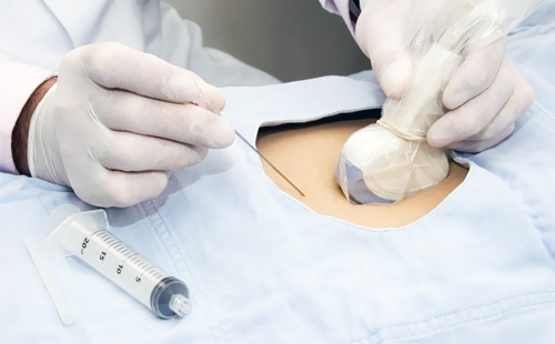
Amniotic Synthesis: With the help of ultrasound imaging, a needle is passed through the pregnant woman's abdominal wall and uterus to the placenta and 10-30 mg is taken from the placenta. It is usually performed in the 9th week of pregnancy to show abnormal symptoms. 15-20 ml of fluid around the baby. This fluid is made up of the baby's urine and cells. This fluid replenishes itself over time. The best time to perform this procedure is 15-20. In some special cases, early amniocentesis or late amniocentesis can be performed, even though there are several weeks of gestation.
Cord Synthesis: This is the process of drawing blood from the baby's umbilical cord. Cord puncture is a procedure performed after the 16th or 18th week of pregnancy to determine whether there are any genetic problems in the unborn baby or infection in the uterus. Since the blood taken from the cord contains the baby's cells, it means that blood is taken from the baby.
The study of chromosomes obtained from peripheral blood, bone marrow, skin, etc. tissue samples by short-term or long-term cell culture is called postnatal cytogenetic diagnosis. Samples taken from patients are analyzed according to their indications and the results are interpreted. Patients receive a report with genetic counseling. The duration of these procedures is usually about 3 weeks


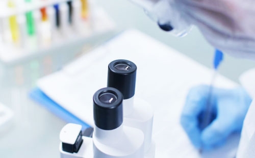


Our center mainly provides services in the fields of genetic counseling, cytogenetics, molecular cytogenetics and molecular genetics. The problems of our patients who apply to our center, which are thought to have genetic causes, are helped by using up-to-date information and technology.
Chromosome disorders form an important category of genetic disease. Cytogenetic tests examine the number and morphology of chromosomes.
Diseases that cause deterioration of human health and change the living order are diagnosed and detected through molecular tests.
It is a discipline that expands the scope and increases the diagnostic value of routine chromosome analysis with the combined use of molecular biology and cytogenetic techniques.
Diagnosis of cancer at an early stage greatly affects the success of cancer treatment. Many cancer tests are performed in our genetic diagnosis center.


We offer genetic analysis and clinical consultancy services at the highest technology and quality standards, using the most up-to-date scientific knowledge, diagnosis and treatment methods.
 Ankara Genetic Diagnosis CenterGenetics Laboratory